Using the Slider Bar
Introduction

|
The slider bar provides a convenient way to scroll through images such as MG tomosynthesis, CT images, or 3D views provided the series has the minimum required images. See your System Administrator to change the minimum required images. Synapse displays orientation markers at each end of the slider bar. You can click an orientation marker to jump to the beginning or the end of a series. Note: The slider bar does not display icons for the following annotations or bookmarked series: 3D quadrant view, All Images, 3D sphere, and Bookmarks. |
Steps
- Select a series that contains the minimum required images. Synapse automatically displays the slider bar; the slider bar cannot be hidden.
- Drag the slider bar control
 to scroll through the images.
 - Optional: Click 2D or 3D
 to access
the slider bar for MG tomosynthesis.
 - Do any of the following:
|
If you want to ... |
Do this ... |
|---|---|
|
See an image with an annotation. |
Click
Annotation Note: The annotation icon is not available for 3D series. |
|
View a bookmark image. |
Click
Bookmark Note: The bookmark icon is not available for 3D series. |
|
Access Images with GSPS annotation overlays. |
Click Presentation State Note: The presentation state icon is not available for 3D series. |
|
View the MG CAD SR finding for a breast tomosynthesis image. |
Click MG CAD Findings |
|
|
When you increase the slab thickness,
the slider bar control becomes larger |
Additional Information
Non-Breast Tomosynthesis Slider Bar Orientation Markers
Synapse supports the following non-breast tomosynthesis modality orientation markers:
- Head (H)
- Foot (F)
- Left (L)
- Right (R)
- Anterior (A)
- Posterior (P)
If patient orientation is not available or the image has a mixed orientation, Synapse displays the first/last image index. The top label is always Image 1 and the bottom label displays the image/frame count of the series.
Breast Tomosynthesis Slider Bar Orientation Markers
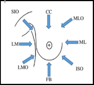
This image provides the mammography views used for all breast images.

The following table lists the breast tomography views and orientation markers, view codes, and slider bar location (top or bottom), and whether it is the first or last image.
|
View |
CC |
FB |
MLO |
LMO |
ML |
LM |
ISO |
SIO |
XCC
|
|||||||||||||||||||||||||||||
|
View
|
R-10242 |
R-10244 |
R-10226 |
R-10230 |
R-10224 |
R-10228 |
R-40AAA |
R-102D0 |
R-102CF
|
|||||||||||||||||||||||||||||
|
|
|
|
|
|
|
|
|
|
|
Note: |
The red orientation markers listed above, represent the bottom slider bar image and Image 1 on the slider bar. |
|---|
 on the slider bar. See
  .
.